Thoracic Endovascular Aortic Repair (TEVAR) is a minimally invasive procedure to repair the major blood vessel in the body, called the aorta.
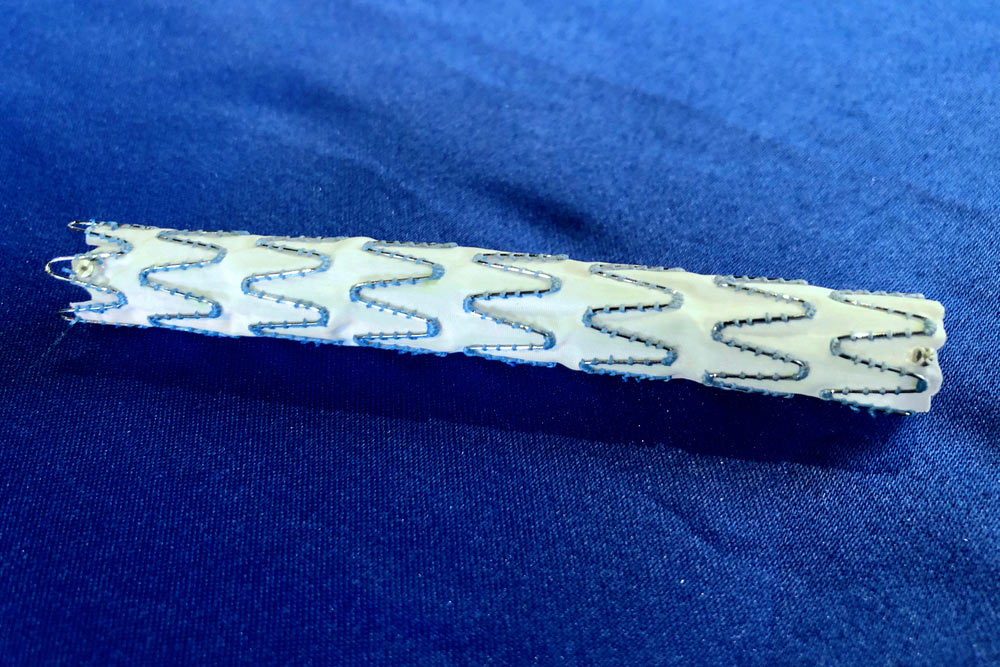
The aorta exits the heart and carries blood to all the organs and the rest of the body. After leaving the heart, the aorta branches to the arms and the brain before running down the back of the chest (thorax) into the belly (abdomen). The aorta forks at the level of the belly button (umbilicus) into branches that go down each leg. To fix or “re-line” the aorta, doctors place a device through a small hole in your groin, known as a stent graft. This device is made of a fabric-covered metal mesh which is fully opened under X-ray. The device repairs the diseased aorta and helps to keep it open and allow blood to flow properly to the rest of the body. The aorta can be affected by a number of different diseases including aneurysm, dissection, transection and stenosis.
- Depending on the type of disease, the TEVAR procedure usually provides a cure. The procedure usually takes around 2 hours to complete.
- The alternative to TEVAR is what doctors refer to as “open repair”. This procedure requires a large incision through the breastbone or side of the chest, much more invasive compared to the small groin incision made with TEVAR.
- Current devices (stent grafts) have a lifespan of at least 10 years.
Why It’s Done
Reasons why you might need TEVAR:
- A weakening of the wall or ballooning out of the aorta, or what doctors call an aneurysm
- Separation of one of the aorta’s three layers, or what doctors call dissection
- Narrowing of the inside of the aorta, or what doctors call stenosis
- Damage to the aorta from trauma or a car accident, or what doctors call transection
Description
- This procedure can be performed under local or general anesthesia.
- Access to the blood vessel is made through a small puncture in the groin.
- A small plastic tube is placed into the aorta to take a picture (angiogram).
- Dye is injected into the small tube, to provide a roadmap of the blood vessels.
- The diseased area of the aorta is marked and measured for placement of the device.
- The device is delivered to the affected area over a wire, which acts like a rail.
- The device is deployed (opened) under the x-ray guidance.
- Dye is injected again to confirm the final position of the device.
- The puncture site in the groin is closed with a plug.
- Possible discomfort or pain
Risks
- Problems at the puncture site in the groin
- Bleeding or infection
- The most severe complications of the procedure are stroke or weakness in the legs (paraplegia)
- Although rare, some very sick patients can also have a heart attack after major surgery
How to Prepare
- Stop smoking.
- Stop blood thinners, per your doctor’s recommendations.
What Can I Expect After Treatment?
- Full return to normal activities
- Monitoring of the stent graft device with annual CT images at your doctor’s office